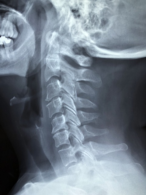
Contrast agents play a crucial role in enhanced X-ray imaging, enabling healthcare professionals to distinguish between tissues and structures within the body. By highlighting areas of interest, these agents improve visualization of tumors, blockages, and abnormalities not visible in standard X-rays. Enhanced X-ray imaging is vital for diagnosing cardiovascular diseases, digestive issues, and bone fractures, leading to more accurate diagnoses, improved treatment outcomes, and better patient care.
“Contrast plays a pivotal role in medical imaging, enabling healthcare professionals to detect tumors, blockages, and abnormalities with unprecedented clarity. This article delves into the intricate world of contrast agents and their synergistic effect with advanced technologies like enhanced X-ray imaging. From understanding the fundamentals of contrast enhancement to exploring its practical applications, we unravel how these innovations are revolutionizing diagnostic precision and patient outcomes.”
Understanding Contrast in Medical Imaging
Contrast plays a pivotal role in medical imaging, enabling healthcare professionals to distinguish between different tissues and structures within the human body. In enhanced X-ray imaging, for instance, contrast agents are introduced into the patient’s bloodstream to highlight specific areas of interest. These agents absorb or reflect X-rays differently than surrounding tissues, creating distinct patterns that can reveal anomalies like tumors, blockages, or abnormalities not readily apparent in standard images.
By enhancing certain structures or organs, medical professionals gain valuable insights into a patient’s health status. This technique is particularly useful in diagnosing conditions such as cardiovascular diseases, digestive issues, and bone fractures. The strategic use of contrast agents and imaging technologies like X-rays ensures more accurate diagnoses, leading to improved treatment outcomes and better patient care.
Enhancing X-ray Technology for Better Visualization
In recent years, enhanced X-ray imaging has emerged as a powerful tool in medical diagnostics, revolutionizing the way healthcare professionals visualize internal structures. By utilizing advanced techniques and computer processing, standard X-rays are transformed into highly detailed images, revealing even the subtlest abnormalities. This technology is particularly beneficial for detecting tumors, blockages, and other anomalies within the human body.
One of the key advantages of enhanced X-ray imaging is its ability to provide clearer, more contrast-rich pictures. Through various methods like digital enhancement, density contrast techniques, and specialized filters, medical experts can improve the visibility of critical areas. This increased contrast allows for more accurate identification and measurement of potential issues, leading to faster and more effective patient care.
Detecting Tumors: The Role of Contrast Agents
Contrast agents play a pivotal role in enhancing X-ray imaging, making it an invaluable tool for detecting tumors and abnormal growths within the body. These agents are substances administered to patients before an X-ray examination, designed to contrast with the surrounding tissue, highlighting specific areas of interest. When incorporated into tissues with differing density, contrast agents absorb or scatter X-rays in unique ways, allowing radiologists to visualize structures that might otherwise be obscured.
For instance, in the case of tumors, which often have a distinct density compared to healthy tissue, contrast agents can provide sharp, clear images, enabling healthcare professionals to accurately identify and assess their size, shape, and location. This early detection is crucial for successful treatment outcomes, as it allows for timely intervention and accurate staging of the cancer.
Uncovering Blockages and Abnormalities with Contrast
In the realm of medical diagnostics, contrast agents play a pivotal role in enhancing X-ray imaging, enabling healthcare professionals to uncover blockages and abnormalities with remarkable clarity. When introduced into the body, these agents reflect X-rays differently than surrounding tissues, creating distinct patterns that stand out against the background. This stark contrast acts as a beacon, highlighting areas where normal flow or structure is obstructed or deviates from the norm.
For instance, in the case of blocked arteries or intestinal obstructions, the contrast agent’s unique interaction with X-rays reveals narrowings or complete blockages, providing critical insights for accurate diagnosis and treatment planning. Similarly, contrasting tissues can highlight tumors or cysts, allowing radiologists to differentiate them from surrounding healthy organs. This enhanced visualization not only aids in early detection but also facilitates more precise interventions, ultimately improving patient outcomes.
Contrast plays a pivotal role in medical imaging, enabling healthcare professionals to visualize tumors, blockages, and abnormalities more clearly. Enhanced X-ray imaging techniques, such as those discussed, harness the power of contrast agents to reveal hidden details that might otherwise go unnoticed. By leveraging these advancements, doctors can make more accurate diagnoses and develop effective treatment plans, ultimately improving patient outcomes.
