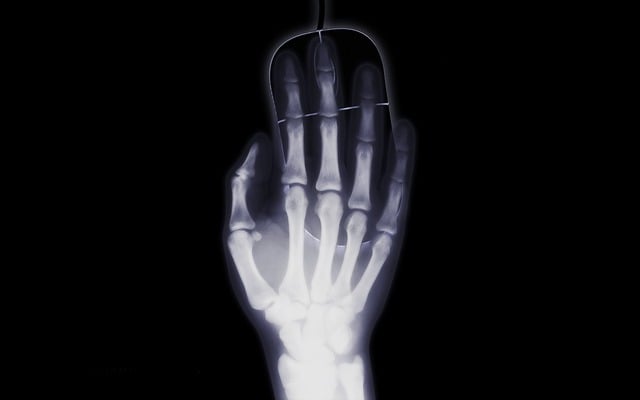
Contrast media are crucial for angiography, enhancing blood vessel visibility and aiding in diagnosing circulatory system conditions. Safe administration involves rigorous testing, precise dosing, patient history review, and monitoring to minimize risks associated with X-ray procedures, ensuring optimal care for patients undergoing imaging.
“Contrast media plays a pivotal role in angiography, enhancing blood vessel visibility for accurate imaging. This article delves into the intricacies of contrast media used in X-ray procedures, exploring its understanding, benefits, and associated risks. We examine the different types of contrast agents and their unique functions, while emphasizing the importance of safe administration practices. By understanding the safety aspects of X-ray contrast media, healthcare providers can ensure optimal imaging outcomes with minimal complications.”
Understanding Contrast Media in Angiography
Contrast media play a pivotal role in angiography, enhancing the visibility of blood vessels and related structures during imaging procedures. These substances are designed to improve the contrast between the vessels and surrounding tissues, allowing radiologists to more accurately diagnose conditions affecting the circulatory system. In angiography, X-ray dyes or contrast agents are introduced into the bloodstream, typically via an IV injection. As these media flow through the vasculature, they temporarily outline the vessel walls, making them easier to identify on X-ray images. This is particularly crucial for detecting blockages, abnormalities, or leaks within the intricate network of blood vessels.
The safety of contrast for X-ray procedures has been a subject of extensive research and improvement. Modern contrast media are formulated to minimize potential risks while maximizing diagnostic benefit. These agents undergo rigorous testing to ensure their compatibility with various bodily systems, including the kidneys and cardiovascular network. Additionally, advanced technologies enable more precise dosing and administration, reducing side effects commonly associated with older contrast materials. Understanding the mechanisms and safety profiles of these media is essential for healthcare professionals conducting angiography and ensuring optimal patient care.
Benefits and Risks of X-ray Contrast Safety
The safety of contrast for X-ray is a critical aspect of angiography and blood vessel imaging procedures. One of the primary benefits of using contrast media is its ability to enhance visibility, allowing radiologists to better visualize the intricate structures within the body’s vasculature. These agents act as X-ray absorbers, creating distinct contrasts between the vessels and surrounding tissues, which aids in detecting abnormalities like blockages or leaks. This improved visibility can lead to more accurate diagnoses and informed treatment decisions.
However, despite its advantages, the safety of contrast for X-ray also involves considerations. As with any medical procedure, there are potential risks associated with the use of contrast media. Common side effects include allergic reactions, though these are relatively rare. More severe complications can occur in patients with kidney issues or other coexisting conditions. Therefore, healthcare providers must carefully assess patient history and conduct thorough screenings before administering contrast agents to ensure safe imaging procedures.
Types of Contrast Agents and Their Functions
Contrast media play a pivotal role in angiography and blood vessel imaging, enhancing visibility and providing crucial diagnostic information. These agents are designed to improve the contrast between the vessels and surrounding tissues on X-ray images. They can be broadly categorized into two types: ionic and non-ionic contrast agents.
Ionic contrast agents, containing metal ions like iodine or barium, are effective in improving image quality due to their high X-ray absorbance. These agents are often used in procedures such as angiography and venography for detailed visualization of blood vessels. Non-ionic contrast agents, on the other hand, do not contain ionizable components, making them safer for patients with kidney issues, a common concern related to the safety of contrast for X-ray imaging. They have become increasingly popular due to their low toxicity and minimal side effects.
Best Practices for Safe Administration
The safe administration of contrast media is paramount in angiography procedures. Healthcare professionals must adhere to best practices to ensure minimal risks and optimal imaging results. This includes meticulous patient preparation, where a thorough medical history review helps identify any potential contraindications or allergies to contrast agents. Additionally, appropriate training for healthcare providers is essential to prevent adverse reactions and accurately interpret imaging findings.
During the procedure, strict adherence to dosage guidelines and injection techniques is crucial. Slow and controlled injections reduce the chances of adverse effects, such as vasospasm or allergic reactions. Continuous monitoring of vital signs also allows for prompt intervention if any complications arise. Furthermore, using high-quality contrast media with well-characterized safety profiles minimizes the risk of adverse events, enhancing the overall safety of X-ray procedures.
Contrast media play a pivotal role in angiography, enhancing the visibility of blood vessels and facilitating accurate diagnosis. By understanding the types of contrast agents, their functions, and adhering to best practices for safe administration, healthcare professionals can maximize the benefits while minimizing risks associated with X-ray contrast safety. This balanced approach ensures effective imaging procedures that rely on these media without compromising patient well-being.
